Choledochal cyst is a rare congenital condition in which the bile ducts become abnormally dilated, leading to abdominal pain, jaundice, digestive problems, and, if left untreated, serious complications such as infection or even liver damage. At Graphic Era Hospital, Dehradun, our team of hepatobiliary and gastrointestinal surgeons performs advanced procedures such as choledochal cyst excision with bile duct reconstruction (hepaticojejunostomy) to restore normal bile drainage and prevent future complications. Using high-definition imaging, minimally invasive laparoscopic techniques, and precise anaesthesia monitoring, we ensure every surgery is performed with maximum safety, causes minimal scarring, and has a fast recovery.
What is a Choledochal Cyst?
A choledochal cyst is an abnormal swelling or dilation in the bile duct – the tube that carries bile from the liver to the small intestine. This condition can occur in both children and adults and often affects the normal flow of bile, leading to symptoms such as abdominal pain or jaundice.
At Graphic Era Hospital, we use advanced radiology imaging, including ultrasound, CT scan, and MRCP, to detect the cyst early and assess its type and extent. Early diagnosis allows our gastrointestinal surgeons to plan the most effective treatment approach and prevent complications such as infection, pancreatitis, or liver damage.

What are the Types of Choledochal Cysts?
Doctors classify choledochal cysts into five main types depending on where the swelling occurs in the bile duct system. Identifying the correct type helps determine the most effective surgical approach. Different types are:
- Type I: The most common form, characterised by a balloon-shaped swelling of the main bile duct located outside the liver.
- Type II: Small pouch (diverticulum) branching out from the bile duct.
- Type III: Cyst located in the section of the bile duct that passes through the intestine.
- Type IV: Multiple cysts affecting bile ducts both inside and outside the liver.
- Type V (Caroli’s Disease): Cysts limited to the bile ducts within the liver.
What Causes a Choledochal Cyst?
The exact cause of choledochal cysts is not always clear, but several factors are known to contribute to their development. In most cases, the condition is congenital, meaning it is present from birth.
Common contributing factors include:
- Abnormal junction of bile and pancreatic ducts, causing backflow of digestive enzymes that weaken the bile duct wall.
- Congenital weakness or malformation of the bile duct structure.
- Genetic predisposition, which may increase the likelihood of cyst formation in some families.
- Chronic bile reflux and inflammation, especially in cases diagnosed later in life.
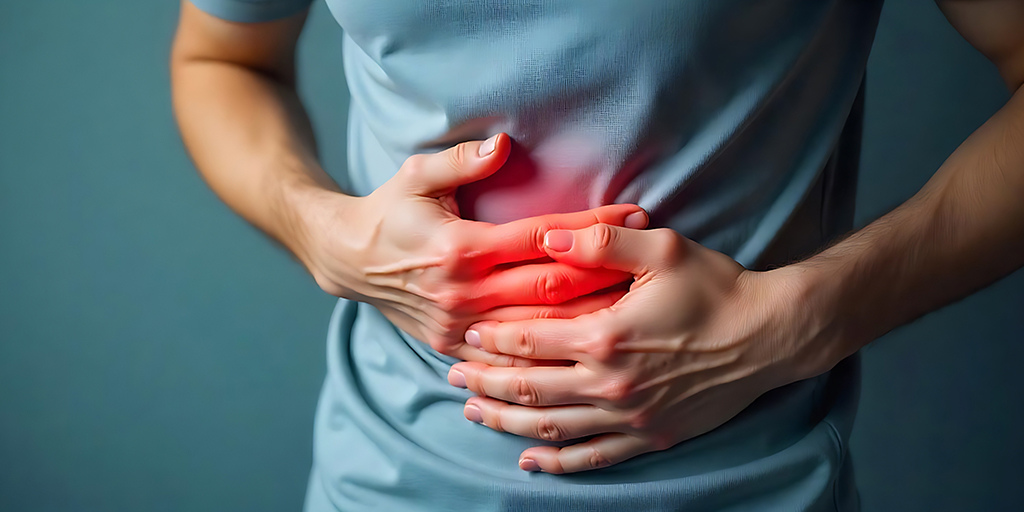
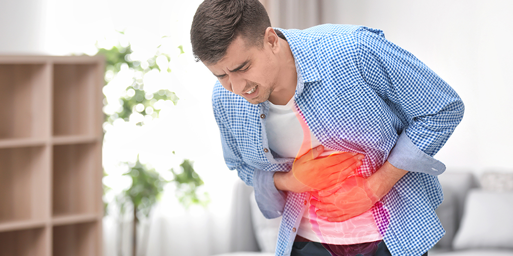
What are the Symptoms of a Choledochal Cyst?
The symptoms of a choledochal cyst vary depending on the patient’s age and the type of cyst, but most are related to obstruction or infection in the bile duct. Early recognition of these signs allows timely treatment and prevents liver complications.
Common symptoms include:
- Persistent abdominal pain or discomfort in the upper right side.
- Jaundice, causing yellowing of the skin and eyes.
- Nausea and vomiting due to bile flow obstruction.
- Fever or chills from bile duct infection (cholangitis).
- Swelling or lump felt in the upper abdomen.
- Clay-coloured stools or dark urine caused by blocked bile flow.
- Pancreatitis, or inflammation of the pancreas, in advanced cases.
How is a Choledochal Cyst Diagnosed?
Accurate diagnosis is essential to determine the type, size, and severity of a choledochal cyst. Our gastroenterologists use advanced imaging and laboratory investigations to confirm the diagnosis and plan the most effective treatment.
Key diagnostic methods include:
- Ultrasound Imaging: Often the first test to detect cystic dilations in the bile duct.
- CT Scan: Provides detailed images of the liver, bile ducts, and surrounding structures.
- Magnetic Resonance Cholangiopancreatography (MRCP): A specialised MRI technique that gives a clear picture of the bile and pancreatic ducts without radiation.
- Blood Tests and Liver Function Tests: Assess bile flow obstruction, inflammation, and liver health.
- Endoscopic Retrograde Cholangiopancreatography (ERCP): Occasionally used to diagnose and treat associated digestive issues.
What is the Treatment for a Choledochal Cyst?
Surgery is the only definitive treatment for a choledochal cyst. Early removal of the cyst prevents serious complications such as infection, pancreatitis, and liver damage.
The standard treatment involves:
- Choledochal Cyst Excision: Complete removal of the cystic portion of the bile duct to eliminate the source of obstruction and infection.
- Hepaticojejunostomy: Reconstruction of the bile duct by connecting it to a part of the small intestine, allowing normal bile flow.
- Laparoscopic or Open Surgery: Surgeons may perform the procedure through minimally invasive laparoscopic techniques or a traditional open approach, depending on the cyst’s size and location.
This comprehensive approach ensures long-term relief, restores bile drainage, and significantly reduces the risk of recurrence or bile duct cancer.
What Happens During Choledochal Cyst Excision?
Choledochal cyst excision is a carefully planned surgical procedure performed under general anaesthesia. Our surgical team follows a structured, safety-focused approach to ensure complete cyst removal and smooth recovery.
The procedure involves the following steps:
- Anaesthesia and Preparation: The patient is placed under general anaesthesia, and the abdomen is prepared for surgery.
- Cyst Removal: The surgeon removes the entire cystic segment of the bile duct, taking care to preserve healthy tissue and surrounding structures.
- Bile Duct Reconstruction: A new connection is created between the bile duct and the small intestine (hepaticojejunostomy) to restore normal bile drainage.
- Minimally Invasive Option: In suitable cases, surgeons perform laparoscopic choledochal cyst excision, which requires smaller incisions and allows faster recovery.
- Closure and Monitoring: The incision is closed, and the patient is closely monitored during recovery to prevent infection and ensure smooth healing.
Why Choose Graphic Era Hospital for Choledochal Cyst Treatment in Dehradun?
Expert Hepatobiliary Surgeons: Our specialists have extensive experience in performing complex bile duct and liver surgeries, including laparoscopic and open choledochal cyst excision.
Advanced Imaging and Operation Theatres: We use high-definition laparoscopic systems and real-time radiology support for accurate diagnosis and precision-guided surgery.
Comprehensive Pre- and Postoperative Care: From detailed evaluation to follow-up rehabilitation, we manage every stage of treatment with precision and care.

Postoperative Care and Recovery
Recovery after choledochal cyst surgery is an important phase where close medical supervision and patient cooperation ensure the best results. Our surgical and nursing teams provide continuous care and guidance throughout this period. Postoperative care includes:
- Hospital Stay: Most patients stay for 5–7 days to allow safe recovery and wound monitoring.
- Diet and Hydration: Patients start with liquids and gradually return to a normal diet as digestion improves.
- Pain and Infection Management: Medications and regular wound checks help reduce discomfort and prevent infection.
- Activity Guidance: Gentle movement is encouraged early to promote circulation, with a gradual return to regular activities over a few weeks.
- Follow-Up Appointments: Scheduled check-ups and imaging tests ensure the reconstructed bile duct is functioning properly.
With proper care, most patients recover fully within a few weeks and regain normal liver and digestive function without long-term complications.
What are the Risks or Complications if Left Untreated?
If a choledochal cyst is not treated in time, it can lead to serious and sometimes irreversible complications. Early surgical management helps prevent these long-term risks.
Possible complications include:
- Recurrent bile duct infections (cholangitis) that cause fever, pain, and liver inflammation.
- Pancreatitis, resulting from bile reflux into the pancreatic duct.
- Liver fibrosis or Liver cirrhosis due to prolonged bile obstruction.
- Stone formation within the bile duct or gallbladder.
- Cyst rupture or leakage, leading to severe infection in the abdomen.
- Increased risk of bile duct cancer (cholangiocarcinoma) if left untreated for several years.
Top Choledochal Cyst Treatments at Graphic Era Hospital
- Laparoscopic Choledochal Cyst Excision
- Open Choledochal Cyst Excision
- Hepaticojejunostomy (Bile Duct Reconstruction)
- Paediatric Choledochal Cyst Surgery
- Revisional Surgery
Top Procedures
- Peroral endoscopic myotomy (POEM)
- EUS-guided Hepaticogastrostomy (EUS-HGS)
- Endoscopic Mucosal Resection (EMR)
- EUS-Guided Gastrojejunostomy
- Endoscopic Submucosal Dissection (ESD)
- Lumen-Apposing Metal Stent (LAMS)
- Full-Thickness Resection Device (FTRD) Procedures
- EUS-Guided Gastric Coil Embolization
- EUS-Guided Antegrade Stenting (EUS-AGS)
- EUS-guided Choledochoduodenostomy (EUS-CDS)
- Metal Stent Placement
- Laparoscopic Cholecystectomy
- Z-POEM (Peroral Endoscopic Myotomy)
- Gallbladder Cancer Surgery (Laparoscopic & Open)
- Gastric Cancer Surgery (Gastrectomy)
- Laparoscopic Sleeve Gastrectomy
- Gastric Bypass Surgery
- Laparoscopic Appendectomy
- Laparoscopic Splenectomy
- Splenorenal Shunt Surgery
Blog


Frequently Asked Questions
What is a choledochal cyst and how is it treated?
A choledochal cyst is a swelling in the bile duct that affects the normal flow of bile. The treatment involves surgical removal of the cyst and reconstruction of the bile duct through a procedure called hepaticojejunostomy.
Is a choledochal cyst serious?
Yes. If left untreated, a choledochal cyst can lead to recurrent infections, liver damage, pancreatitis, or even bile duct cancer. Early surgical intervention ensures long-term recovery and prevents complications.
What causes choledochal cysts in children?
Most cases are congenital, meaning they are present at birth. The condition often results from an abnormal connection between the bile and pancreatic ducts, which causes bile reflux and duct wall weakening.
How is a choledochal cyst diagnosed?
Diagnosis is done through imaging tests such as ultrasound, CT scan, or MRCP, which help identify the cyst’s size, type, and exact location.
What is choledochal cyst excision surgery?
It is a surgical procedure where the cystic portion of the bile duct is removed, and a new bile passage is created by connecting the bile duct to the intestine for normal drainage.
How long does recovery take after choledochal cyst removal?
Most patients recover within 4–6 weeks, depending on the type of surgery performed and overall health. Laparoscopic excision usually offers a faster recovery.
Is laparoscopic surgery possible for choledochal cysts?
Yes, many patients are suitable candidates for laparoscopic choledochal cyst excision, which involves smaller incisions, less pain, and quicker recovery than open surgery.
Can a choledochal cyst recur after surgery?
Recurrence is rare when the cyst is completely removed. Regular follow-up and imaging tests help ensure long-term success.
What is hepaticojejunostomy and why is it done?
Hepaticojejunostomy is the process of connecting the bile duct to the small intestine after cyst removal to restore normal bile flow and prevent blockage.
How much does choledochal cyst surgery cost in Dehradun?
The cost varies based on the type of surgery and hospital stay. For details or cost estimates, contact Graphic Era Hospital, Dehradun, at 1800-889-7351.
Where can I find choledochal cyst treatment near me in Dehradun?
You can visit the Department of Hepatobiliary and Gastrointestinal Surgery at Graphic Era Hospital, where our expert surgeons offer advanced treatment for both children and adults using minimally invasive techniques.