Hydronephrosis can quietly affect kidney function by causing a build-up of urine, often without clear symptoms in the early stages. As the condition progresses, it may lead to flank pain, recurrent urinary infections, and increasing pressure on the kidneys, making timely evaluation important to prevent long-term damage.
At Graphic Era Hospital in Dehradun, hydronephrosis is assessed through a structured, cause-focused approach rather than managed in isolation. Our specialists identify the underlying reason, whether it is kidney stones, ureteric obstruction, reflux, or external compression, using advanced imaging and diagnostic tools. Treatment is then tailored to relieve the obstruction, preserve kidney function, and reduce the risk of recurrence, ensuring each patient receives timely and appropriate care.
What is Hydronephrosis?
Hydronephrosis is a condition in which one or both kidneys become swollen because urine is unable to drain properly into the bladder. This happens when there is a blockage in the urinary tract, backward flow of urine, or another problem that interferes with normal urine movement. Rather than being a disease on its own, hydronephrosis is usually a sign of an underlying issue that needs timely medical evaluation.
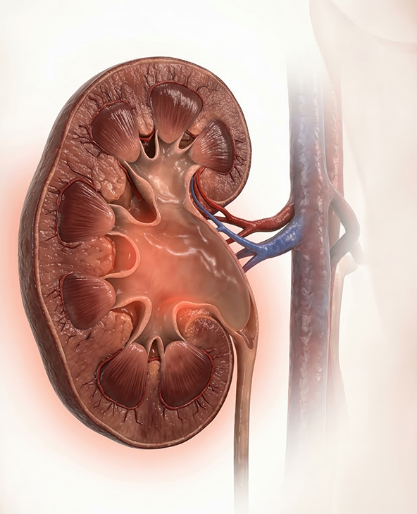
The condition can affect people of all age groups, including infants, children, adults, and pregnant women. In some cases, hydronephrosis causes noticeable symptoms such as pain, fever, or difficulty passing urine, while in others it may be found incidentally during an ultrasound or other imaging test. At Graphic Era Hospital, Dehradun, hydronephrosis is assessed based on its severity, cause, and the extent to which kidney function may be affected, so that treatment can be planned appropriately.
Hydronephrosis is broadly classified based on its severity, extent, and underlying cause:
Mild Hydronephrosis
A slight dilation of the kidney with minimal pressure on kidney structures. It may not cause noticeable symptoms and is often detected incidentally during imaging. Many mild cases can be monitored with regular follow-up.
Moderate Hydronephrosis
Increased swelling of the kidney with more evident pressure on the renal tissue. Patients may begin to experience symptoms such as discomfort, urinary issues, or recurrent infections, requiring closer evaluation and treatment.
Severe Hydronephrosis
Significant dilation of the kidney with thinning of kidney tissue, indicating prolonged pressure and risk of permanent damage. This stage requires prompt intervention to relieve obstruction and preserve kidney function.
Hydronephrosis can also be described in other ways depending on its presentation:
- Unilateral or Bilateral: It may affect one kidney or both, depending on the location of the blockage.
- Acute or Chronic: It can develop suddenly due to conditions such as kidney stones or gradually over time due to long-standing obstruction.
- Obstructive or Reflux-Related: It may result from a physical blockage in the urinary tract or from backward flow of urine from the bladder towards the kidneys.
In children and infants, hydronephrosis may be detected before birth or during early childhood and requires age-appropriate evaluation and monitoring.
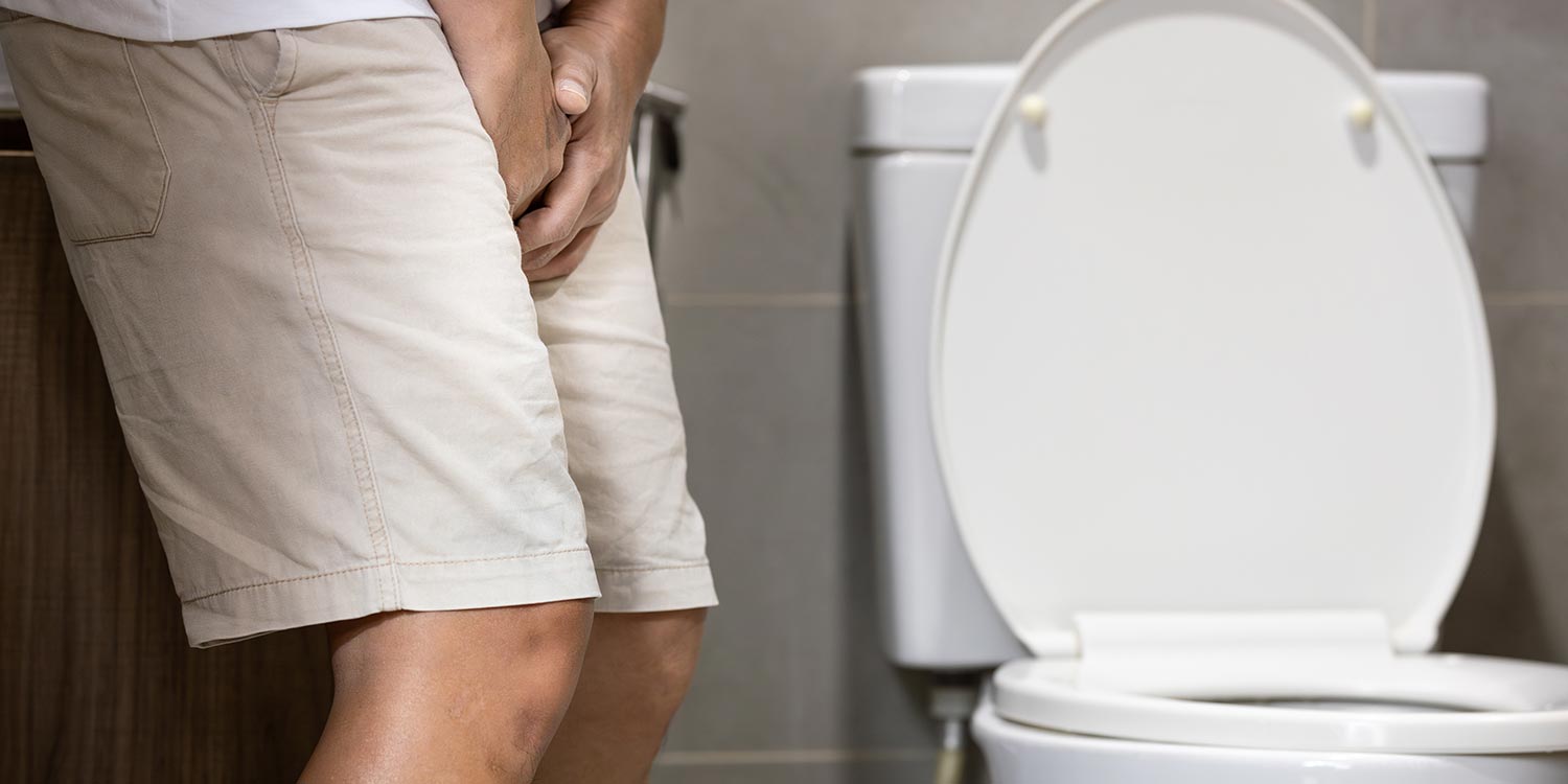
Symptoms of Hydronephrosis and When to See a Doctor
Hydronephrosis can present with a wide range of symptoms depending on its cause, severity, and how quickly it develops. In some cases, especially when the condition develops gradually, there may be no obvious symptoms and it may only be detected during imaging. In other cases, symptoms can be sudden and severe, particularly when there is an acute blockage.
Common symptoms include:
- Flank or Back Pain: Pain on one or both sides of the lower back is one of the most frequent symptoms. It may be dull and persistent in chronic cases or sharp and severe when caused by a sudden obstruction such as a kidney stone.
- Reduced Urine Output: In more severe cases, urine flow may be reduced, particularly when both kidneys are affected or when there is significant obstruction.
- Nausea and Vomiting: These symptoms are often seen in acute hydronephrosis, especially when associated with kidney stones or severe pain.
- Fever and Chills: Fever may indicate an associated urinary tract infection, which can become serious if not treated promptly.
When to Seek Medical Attention
While mild cases may not cause immediate concern, certain symptoms require prompt medical evaluation:
- Severe or worsening flank pain
- Fever accompanied by urinary symptoms
- Noticeable decrease in urine output
- Persistent nausea or vomiting
- Recurrent urinary tract infections
- Symptoms that do not improve or continue to worsen
Early evaluation helps identify the underlying cause and prevents complications such as kidney damage or infection spread.
What Causes Hydronephrosis?
Hydronephrosis usually occurs when urine cannot flow properly from the kidney, causing it to build up and lead to swelling over time. This can happen due to a variety of conditions affecting the urinary tract, some temporary and others requiring medical treatment. Common causes include:
- Kidney Stones: Stones can block the ureter and prevent urine from draining properly. This is one of the most frequent causes of sudden or acute hydronephrosis and often presents with severe flank pain.
- Ureteric Stricture or Narrowing: Scarring or narrowing of the ureter can restrict urine flow over time. This may develop after infections, previous surgeries, or inflammation, and can lead to chronic hydronephrosis if not addressed.
- Enlarged Prostate (Benign Prostatic Hyperplasia): In men, an enlarged prostate can compress the urethra and obstruct urine outflow from the bladder, causing back pressure that affects the kidneys.
- Tumours or Masses: Growths within or around the urinary tract, including tumours of the kidney, ureter, bladder, or nearby organs, can press on the urinary passages and block urine flow.
- Vesicoureteral Reflux (VUR): A condition in which urine flows backwards from the bladder towards the kidneys. This is more commonly seen in children and increases the risk of recurrent infections and kidney swelling.
- Pregnancy: During pregnancy, the growing uterus can press on the ureters and slow down urine flow, leading to temporary hydronephrosis. This is usually monitored and resolved after delivery.
- Neurogenic Bladder: Nerve-related problems affecting bladder control can interfere with proper emptying of the bladder, resulting in urine retention and back pressure on the kidneys.
- Blood Clots or Debris in the Urinary Tract: In some cases, clots or tissue fragments can obstruct urine flow, particularly after trauma, surgery, or certain medical conditions.
- Congenital Abnormalities: Some individuals are born with structural issues in the urinary tract, such as ureteropelvic junction obstruction, which can lead to hydronephrosis from an early age.
- Ignoring the Urge to Urinate or Chronic Retention: Long-standing bladder retention can increase pressure within the urinary system and contribute to hydronephrosis over time.
Doctors Available

Why Choose Graphic Era Hospital, Dehradun, for Hydronephrosis Treatment
When it comes to urological care, Graphic Era Hospital in Dehradun is recognised for its clinical expertise and comprehensive approach. Here’s why patients and their families trust us:
Specialist-Led, Cause-Focused Care : Hydronephrosis is evaluated by experienced urologists and nephrologists who focus on identifying the exact cause rather than managing it as an isolated condition. Each patient is assessed based on severity, underlying issue, and kidney function to ensure the most appropriate treatment plan.
Complete Diagnostic Capability Under One Roof : Accurate diagnosis is essential in hydronephrosis. From ultrasound and CT scans to specialised functional tests, all required investigations are available within the hospital, allowing a seamless and timely diagnostic process without delays.
Advanced and Minimally Invasive Treatment Options : Treatment is tailored to the underlying cause and may include minimally invasive procedures such as stenting, endoscopic stone removal, or corrective surgery where needed. The focus remains on relieving obstruction, preserving kidney function, and supporting faster recovery.

Hydronephrosis Care at Graphic Era Hospital: From Diagnosis to Treatment
Diagnostic Approach
Accurate diagnosis is essential in hydronephrosis to determine the cause, severity, and impact on kidney function. At Graphic Era Hospital, evaluation follows a structured approach to identify the exact site of obstruction and guide the most appropriate treatment plan. The assessment typically includes:
- Clinical History and Physical Examination: A detailed review of symptoms such as pain, urinary changes, infections, and past medical history helps identify possible causes. Examination may reveal tenderness or signs of urinary retention.
- Urine Tests: Urine analysis helps detect infection, blood in urine, or other abnormalities that may indicate underlying urinary tract issues.
- Blood Tests: Tests such as serum creatinine and urea are used to assess kidney function and determine whether hydronephrosis has affected the kidneys.
- Ultrasound (USG): Ultrasound is usually the first-line investigation. It helps detect kidney swelling, assess severity, and identify common causes such as stones or obstruction.
- CT Scan: A CT scan provides detailed imaging of the urinary tract and is especially useful in identifying kidney stones, strictures, tumours, or other structural causes.
- CT Urography or IVP: These imaging studies help evaluate the flow of urine and pinpoint the exact location of blockage.
- Nuclear Renal Scan: This specialised test assesses how well each kidney is functioning and how effectively urine is draining, particularly in chronic or complex cases.
Treatment Approach
Treatment for hydronephrosis is guided by the underlying cause, severity of the condition, and the patient’s overall health. The focus is on relieving the obstruction, restoring normal urine flow, and protecting kidney function.
- Monitoring and Observation: Mild cases without significant symptoms or kidney damage may be monitored with regular follow-up and imaging.
- Medical Management: Medications may be used to manage pain or treat associated infections. Antibiotics are prescribed when urinary tract infection is present.
- Ureteric Stenting: A thin tube is placed inside the ureter to allow urine to bypass the blockage and drain properly from the kidney to the bladder.
- Percutaneous Nephrostomy: In more severe cases, a tube may be placed directly into the kidney through the skin to drain urine and relieve pressure.
- Endoscopic Stone Removal (URS / PCNL): When kidney stones are the cause, minimally invasive procedures are used to remove or break the stones and restore urine flow.
- Surgical Correction: Surgery may be required to treat structural problems such as ureteric strictures, congenital narrowing, or tumours causing obstruction.
This structured approach ensures that each patient receives timely, targeted treatment aimed at resolving the cause and preventing long-term complications.
Hydronephrosis in Children and Infants
Hydronephrosis in children and infants requires a different approach from adult care, as it is often detected early, sometimes even before birth during routine pregnancy scans. In many cases, the condition may resolve on its own as the child grows, but some children require careful monitoring or treatment to prevent complications and protect kidney function.
- Antenatal (Before Birth) Detection: Hydronephrosis is commonly identified during prenatal ultrasound. After birth, follow-up imaging is done to assess whether the swelling persists and to determine its cause.
- Monitoring and Follow-Up: Mild cases are often monitored with regular ultrasound scans to track kidney growth and urine flow. Many children improve without the need for intervention.
- Identifying the Underlying Cause: Common causes in children include vesicoureteral reflux, ureteropelvic junction obstruction, or structural abnormalities present from birth. Early evaluation helps guide appropriate management.
- Safe and Age-Appropriate Treatment: When treatment is needed, it is carefully planned based on the child’s age, symptoms, and severity of the condition. Options may include medication, minimally invasive procedures, or surgery in selected cases.
- Parental Guidance and Warning Signs: Parents are advised to watch for symptoms such as fever, poor feeding, irritability, vomiting, or urinary infections. Early medical attention helps prevent complications and supports healthy kidney development.
Top Hydronephrosis Investigations and Treatments
- Ureteric Stenting
- Percutaneous Nephrostomy
- Endoscopic Stone Removal (URS / PCNL)
- Pyeloplasty (for Ureteropelvic Junction Obstruction)
- Surgical Correction of Ureteric Strictures
- Antibiotic Therapy (for Associated Infections)
- Pain Management and Supportive Car
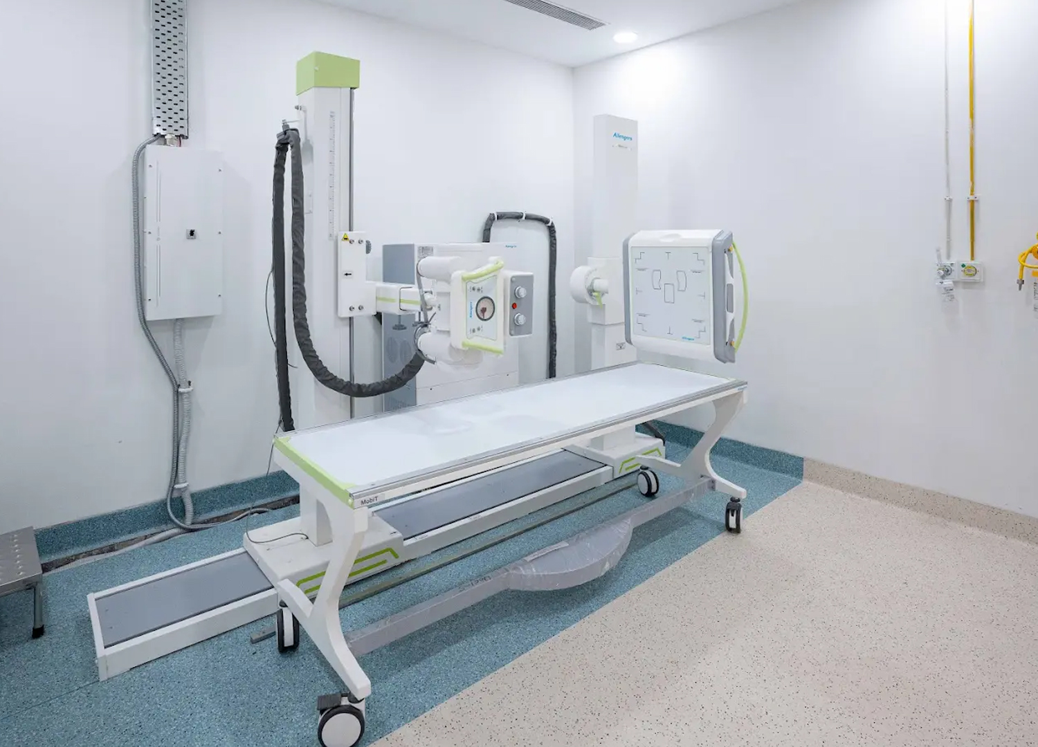
Advanced Diagnostics & Technology
- Offers high-resolution imaging for detailed blood vessel analysis, aiding in accurate diagnosis and treatment planning.
- Delivers advanced imaging with high resolution for clear, detailed views of soft tissues, ensuring precise diagnostics.
- Provides high-quality, detailed radiographic images for accurate diagnosis with minimal exposure to radiation.


Other Specialities
Patient Stories


Blog


Frequently Asked Questions (FAQs)
Can hydronephrosis go away on its own?
In mild cases, especially in children or during pregnancy, hydronephrosis may resolve on its own with monitoring. However, when caused by a blockage or underlying condition, treatment is usually required to prevent complications.
Is hydronephrosis a serious condition?
It can become serious if left untreated. Persistent pressure on the kidney may lead to reduced kidney function or permanent damage, which is why timely evaluation is important.
Does hydronephrosis always require surgery?
No. Treatment depends on the cause and severity. Some cases can be managed with observation or medication, while others may require minimally invasive procedures or surgery to relieve obstruction.
What are the early warning signs of hydronephrosis?
Early signs may include flank or back pain, changes in urination, or recurrent urinary tract infections. In some cases, there may be no symptoms, and the condition is detected during imaging.
How is hydronephrosis diagnosed?
Diagnosis is based on clinical evaluation along with imaging tests such as ultrasound, CT scan, or specialised studies that assess kidney function and urine flow.
Can hydronephrosis affect both kidneys?
Yes. Hydronephrosis can affect one or both kidneys depending on the location and severity of the obstruction. Bilateral involvement requires prompt evaluation.
When should I see a doctor for hydronephrosis?
You should seek medical attention if you experience severe or persistent pain, fever with urinary symptoms, reduced urine output, or recurrent infections.
How do I book an appointment at Graphic Era Hospital?
You can book an appointment through the hospital’s website, call 1800-889-7351, or visit the outpatient department. The team will assist you in scheduling your consultation at the earliest available time.