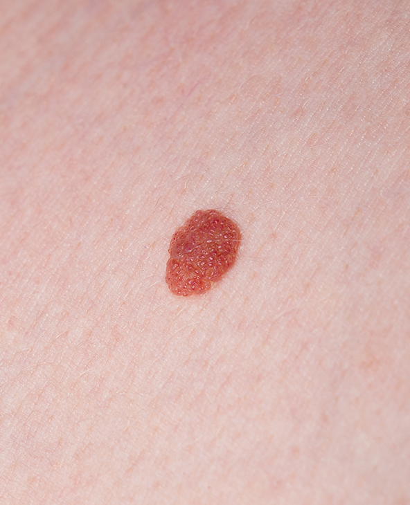
Hemangiomas, also known as infantile hemangiomas, are benign (non-cancerous) growths of blood vessels that typically appear as bright red or bluish marks on a baby’s skin. They may look like a soft, raised bump or a flat patch and commonly develop on the face, scalp, chest, or back, although they can occur anywhere on the body or, less commonly, within internal organs. These growths usually become noticeable within the first few weeks of birth, grow for a period, and then gradually shrink over time, often resolving on their own without treatment.
While many hemangiomas do not require intervention, treatment may be considered if the growth is large, rapidly increasing in size, affecting vital functions such as vision or breathing, or located in cosmetically sensitive areas. At Graphic Era Hospital, Dehradun, hemangioma treatment is guided by careful clinical assessment and a multidisciplinary approach. Our team of highly experienced dermatologists, paediatric specialists, interventional radiologists, and surgeons works together to determine whether active treatment or observation is most appropriate. Each treatment plan is carefully tailored to the child’s condition, with a focus on safety, comfort, and long-term outcomes.
When is Hemangioma Treatment Recommended and Why is it Done?
Hemangioma treatment is not always necessary, as many lesions, especially in infants, may shrink over time. However, intervention is recommended when the growth causes functional impairment, complications, or significant cosmetic concerns. Early evaluation helps prevent progression and ensures timely management when required.
Usually, treatment is advised in the following situations:
- Rapidly growing hemangioma, especially in infants
- Lesions interfering with vision, breathing, feeding, or hearing
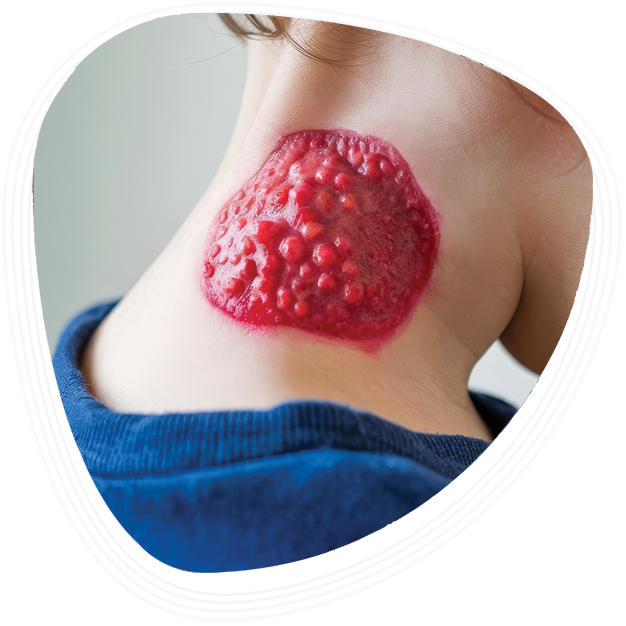
- Ulcerated or bleeding hemangiomas
- Painful or infected lesions
- Hemangiomas located near vital structures (eyes, airway, liver)
- Large or disfiguring facial hemangiomas
- Internal hemangiomas causing symptoms
- Residual skin changes or scarring after involution
- Diagnostic uncertainty requiring further evaluation
Things to Know Before Starting Hemangioma Treatment
Before starting hemangioma treatment, it is important to understand that the approach varies depending on the type (infantile or congenital), depth, location, and growth phase of the lesion. At Graphic Era Hospital, treatment plans are carefully individualised to ensure effective outcomes while maintaining safety, particularly in paediatric cases.
- Not all hemangiomas require active treatment; some are managed with observation
- Infantile hemangiomas typically have a growth phase followed by natural regression
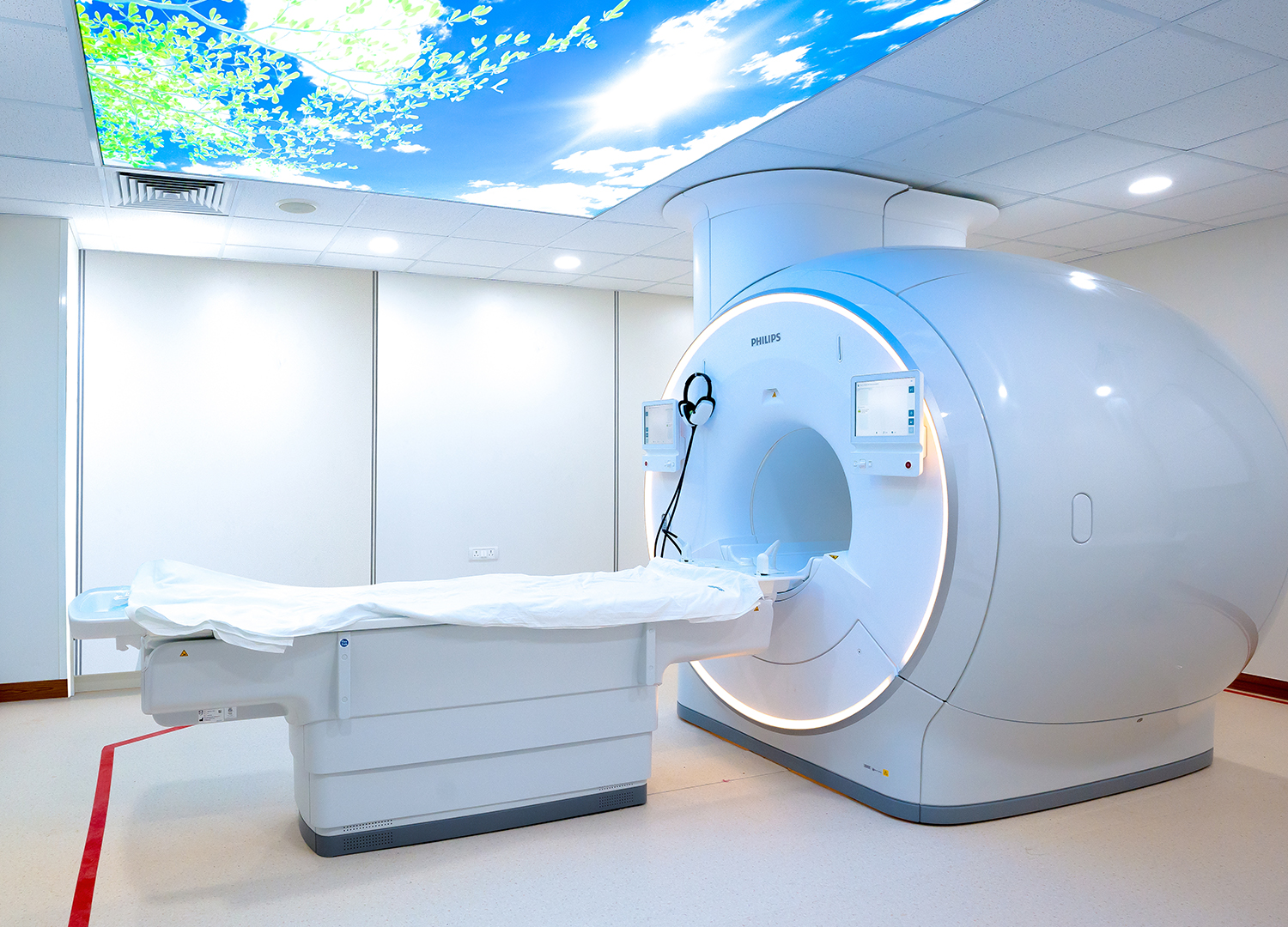
- Imaging such as ultrasound or MRI may be required for deeper lesions
- Treatment choice depends on size, location, and associated complications
- Medical therapy is often the first-line approach for problematic hemangiomas
- Minimally invasive procedures are preferred whenever feasible
- Surgical removal is considered in selected cases
- Cosmetic outcomes are an important part of treatment planning
- Regular monitoring is essential to assess response to treatment
- Long-term follow-up may be required, especially in complex cases
Types of Hemangioma Treatment Available at Graphic Era Hospital
Hemangioma treatment is customised based on whether the lesion is superficial, deep, or involving internal organs. At Graphic Era Hospital, care ranges from observation and medical therapy to laser treatment and surgical intervention, depending on clinical need.
Medical (Non-Surgical) Treatment
- Beta-Blocker Therapy (Propranolol): First-line treatment for many infantile hemangiomas; helps shrink the lesion and slow its growth
- Topical Beta-Blockers (Timolol): Used for small, superficial hemangiomas
- Corticosteroid Therapy: Prescribed in select cases where beta-blockers are not suitable
- Pain and Infection Management: Supportive treatment for ulcerated or complicated lesions
Minimally Invasive & Laser Treatment
- Laser Therapy (Pulsed Dye Laser): Used to reduce redness, treat superficial lesions, or manage ulceration
- Sclerotherapy: Injection-based treatment to shrink certain types of vascular lesions
- Image-Guided Procedures: Minimally invasive techniques for deeper or complex hemangiomas
Surgical Treatment
- Excision Surgery: Removal of residual or problematic hemangiomas, especially when they do not regress completely
- Reconstructive Procedures: Performed when necessary to restore appearance and function after hemangioma removal
Doctors Available

Prof. Dr. Rupa Dalmia Singh
Senior Consultant & HOD
Paediatrics
Experience: 27 Years
Book An Appointment


Dr. Deep Shikha Baranwal
Consultant
Paediatric, Paediatric Nephrology
Experience: 7 Years
Book An Appointment

Why Choose Graphic Era Hospital, Dehradun for Hemangioma Treatment
Hemangioma management requires careful evaluation to determine whether treatment is necessary and, if so, which approach is most appropriate. At Graphic Era Hospital, care is guided by accurate assessment, multidisciplinary expertise, and a strong focus on safety and long-term outcomes, particularly in paediatric patients. We offer:
Specialised Multidisciplinary Expertise : Our dermatologists, paediatric specialists, interventional radiologists, and surgeons work closely to evaluate each case in detail. Treatment decisions are based on the type of hemangioma, its growth phase, anatomical location, and potential functional impact, ensuring that each patient receives appropriately tailored care rather than a one-size-fits-all approach.
Advanced Diagnostic & Imaging Support : Accurate evaluation is essential, particularly for deeper or complex hemangiomas. The hospital utilises imaging modalities such as ultrasound and MRI, along with clinical assessment, to determine lesion depth, vascular involvement, and progression. This enables precise treatment planning and helps avoid unnecessary interventions.
Focus on Safe, Individualised, and Minimally Invasive Care : Whenever possible, non-surgical and minimally invasive treatments such as beta-blocker therapy or laser procedures are prioritised to reduce risk and support natural regression. Surgical intervention is reserved for selected cases, with careful planning to ensure optimal functional and cosmetic outcomes.
Hemangioma Diagnosis & Evaluation at Graphic Era Hospital
Accurate diagnosis is essential to differentiate hemangiomas from other vascular anomalies and to determine whether treatment is required. At Graphic Era Hospital, evaluation combines clinical expertise with advanced imaging.
The diagnostic pathway includes:
- Clinical Examination: Assessment of size, location, and growth pattern
- Ultrasound with Doppler: Evaluates blood flow and lesion characteristics
- MRI Scan: Provides detailed imaging for deep or complex hemangiomas
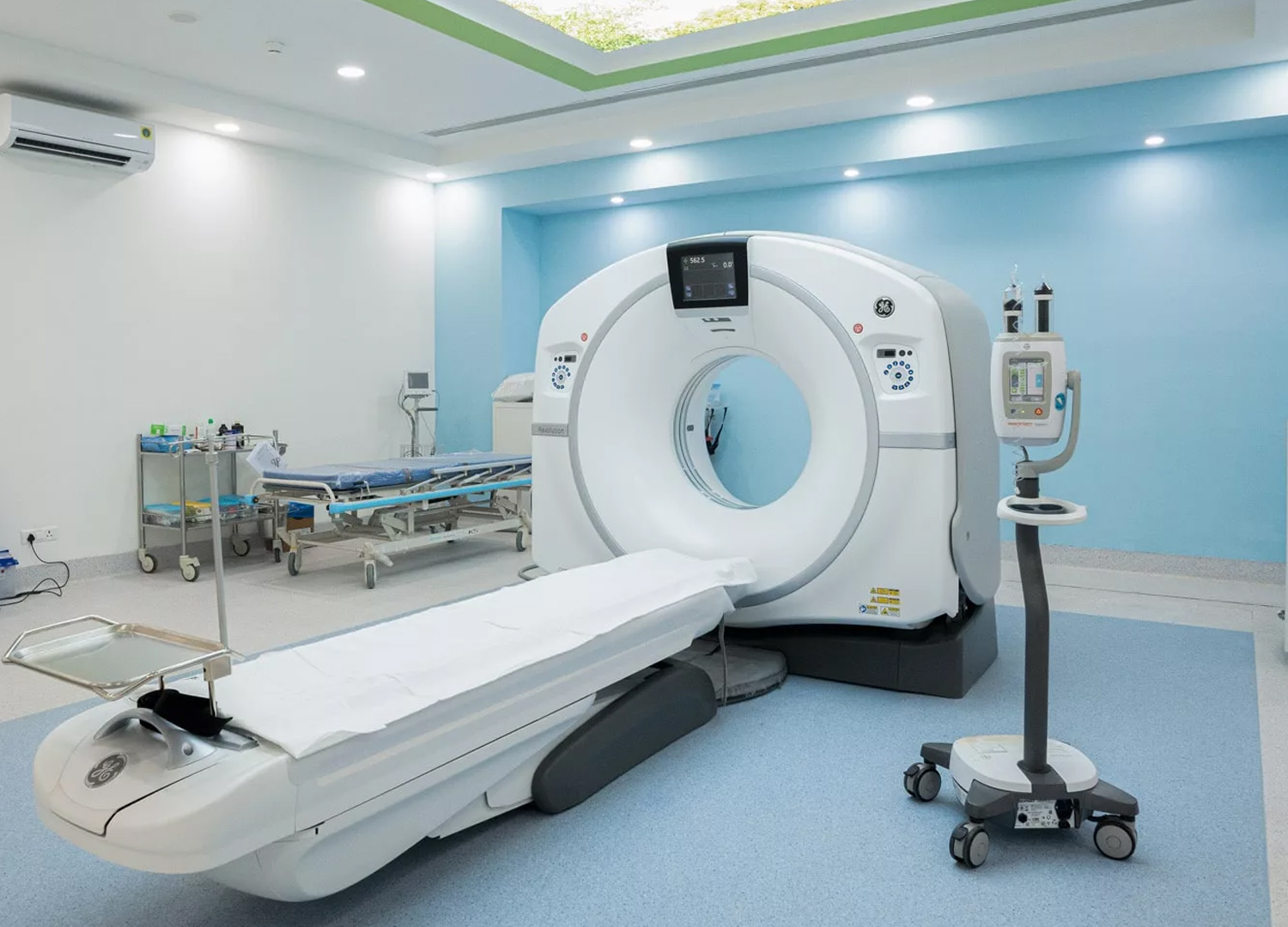
- CT Scan (when required): Used in specific cases involving internal organs
- Biopsy (rare cases): Performed only when diagnosis is uncertain
Hemangioma Recovery & Follow-Up Care
Management of hemangioma extends beyond initial treatment. Regular follow-up is essential to monitor regression, assess treatment response, and address any residual concerns.
Post-treatment care includes:
- Regular Monitoring: Tracking changes in size, colour, and symptoms
- Medication Follow-Up: Adjusting dosage and duration of therapy as needed
- Wound Care: For ulcerated or treated lesions
- Laser Therapy Follow-Up: Multiple sessions may be required for optimal results
- Cosmetic Assessment: Addressing residual marks or skin changes
- Parental Guidance (Paediatric Cases): Educating families on home care and warning signs
- Long-Term Surveillance: Ensuring no recurrence or complications
Top Hemangioma Treatments at Graphic Era Hospital
- Beta-blocker therapy for infantile hemangioma
- Topical treatment for superficial lesions
- Laser therapy for vascular lesions
- Sclerotherapy for selected cases
- Surgical excision and reconstruction
Advanced Diagnostics & Technology
- Offers high-resolution imaging for detailed blood vessel analysis, aiding in accurate diagnosis and treatment planning.
- Delivers advanced imaging with high resolution for clear, detailed views of soft tissues, ensuring precise diagnostics.
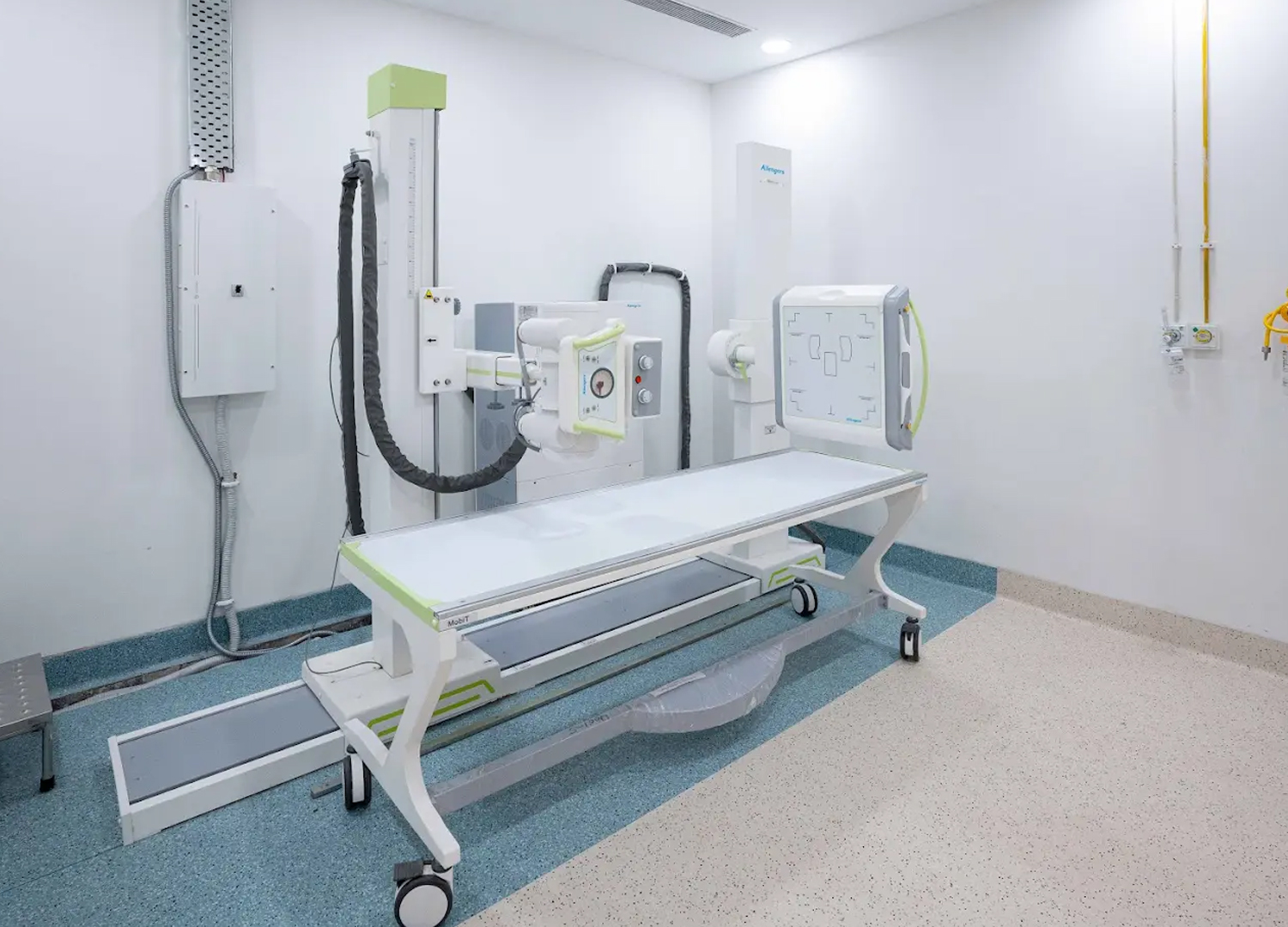
- Provides high-quality, detailed radiographic images for accurate diagnosis with minimal exposure to radiation.
Other Specialities
Patient Stories


Blog


Frequently Asked Questions (FAQs)
Do all hemangiomas require treatment?
No. Many hemangiomas, especially in infants, resolve naturally over time. Treatment is only recommended if complications or functional issues arise.
At what age do hemangiomas usually appear?
Infantile hemangiomas typically appear within the first few weeks of life and may grow during the first year before gradually shrinking.
Is hemangioma a type of cancer?
No. Hemangiomas are benign vascular growths and are not cancerous.
Can hemangiomas become dangerous?
Most are harmless, but those affecting vital areas like the eyes, airway, or internal organs may require prompt treatment.
How long does hemangioma treatment take?
The duration varies depending on the type and severity. Some respond within months, while others may require longer follow-up.
Will a hemangioma leave a scar?
Some may leave mild skin changes or scarring after regression, especially larger lesions. Treatments can help improve cosmetic outcomes.
What are the common hemangioma symptoms?
Hemangioma symptoms vary depending on the type and location of the lesion. Superficial hemangiomas often appear as red or bluish patches or raised bumps on the skin, while deeper lesions may present as swelling beneath the skin. In some cases, symptoms may include rapid growth, ulceration, bleeding, or pain. Hemangiomas located near vital structures may also cause functional issues such as difficulty with vision, breathing, or feeding.
What are the causes of hemangioma?
The exact causes of hemangioma are not fully understood, but they are believed to result from abnormal growth of blood vessels during early development. Infantile hemangiomas are thought to form due to changes in vascular formation before or shortly after birth. Certain factors such as premature birth, low birth weight, and female gender have been associated with a higher likelihood of developing hemangiomas.
Is hemangioma considered a vascular tumour, and how is vascular tumor treatment approached?
Yes, hemangiomas are classified as benign vascular tumours, meaning they arise from an overgrowth of blood vessels. Vascular tumor treatment depends on the type, size, location, and symptoms associated with the lesion. In many cases, observation is sufficient. However, when intervention is required, treatment may include medical therapy such as beta-blockers, laser therapy, minimally invasive procedures, or surgery, depending on the clinical situation.